
In this article (14 sections)1,139 words
- Abstract
- Clinical Bottlenecks & Engineering Objectives
- Core R&D Milestones
- Data Engineering & Expert Clinical Annotation
- Model Selection: Benchmarking Mask R-CNN vs. YOLOv11-seg
- Phase 1: Two-Stage Baseline with Mask R-CNN (MMDetection)
- Phase 2: Transitioning to Single-Stage YOLOv11x-seg (Ultralytics)
- Empirical Benchmark Summary
- Beyond Detection: Deterministic Clinical Intelligence
- 1. Automated Radiographic Bone Loss (RBL %) & AAP 2018 Staging
- 2. Zero-Click 18-Series FMX Periapical Auto-Sequencing
- 3. Rule-Based Treatment Protocol Drafting
- Production Deployment & Infrastructure
- Conclusion & What’s Next
Abstract
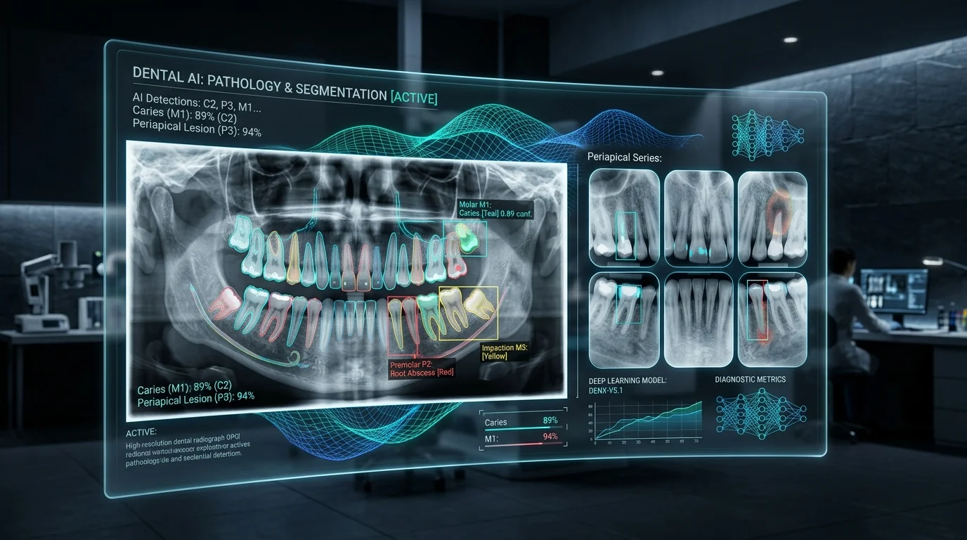
Over the past six months, our engineering and clinical research team embarked on an intensive R&D journey: building a production-grade deep learning engine for dental radiographic diagnostics.
Panoramic radiographs (OPG) and periapical (PA) series remain the diagnostic cornerstone of modern dentistry. While these images contain critical signals for early caries, apical periodontitis, and alveolar bone degradation, manual interpretation places an immense cognitive burden on clinicians. Reading dozens of full-mouth scans daily often leads to diagnostic fatigue and high inter-examiner variability.
Here, we share our technical roadmap, model benchmarking journey from Mask R-CNN to YOLOv11-seg, clinical calculation engines for AAP 2018 periodontal staging, and our production deployment architecture.
Clinical Bottlenecks & Engineering Objectives
In clinical radiology workflows, practitioners face three primary challenges:
- High Cognitive Workload & Diagnostic Fatigue: Interpreting high-resolution radiographs across 32 individual tooth positions—each with complex overlapping structures—is time-intensive.
- Inter-Examiner Variability: Clinical studies demonstrate significant variance between practitioners when identifying early-stage enamel demineralization or subtle apical radiolucencies.
- Subtle & Occult Lesions: Incipient interproximal caries, early furcation involvement, and minor crestal bone resorption are frequently overlooked during routine recall exams.
Core R&D Milestones
To address these pain points, our engineering objectives were defined around three pillars:
- Sub-30ms Real-Time Inference: Deliver instantaneous diagnostic overlays without disrupting the chairside workflow.
- Sub-Millimeter Instance Segmentation: Delineate precise tooth anatomy, pulp chambers, restorations, and pathological boundaries rather than coarse bounding boxes.
- Deterministic Clinical Translation: Automatically compute radiographic bone loss percentages (RBL %) and map findings to standardized diagnostic frameworks (e.g., AAP/EFP 2018 Periodontitis Staging).
Data Engineering & Expert Clinical Annotation
High-quality, specialized ground truth data is the bedrock of medical computer vision. We partnered with clinical dental practices to curate and label thousands of high-fidelity panoramic and periapical X-rays.
To ensure clinical validity, our annotation taxonomy was constructed in collaboration with senior dental specialists across three functional layers:
- Anatomical & Landmark Labels: Individual tooth segmentation with FDI/Universal numbering, enamel-dentin junctions, and root apexes.
- Pathological & Condition Labels: Enamel/dentin caries, periapical lesions, impacted teeth, calculus, and retained root fragments.
- Periodontal Reference Boundaries: The Cementoenamel Junction (CEJ) and Periodontal Bone Level (PBL).
Model Selection: Benchmarking Mask R-CNN vs. YOLOv11-seg
Designing an effective computer vision architecture for dental X-rays requires balancing spatial resolution, small-object detection accuracy, and runtime latency.
[Raw Dental X-Ray (OPG / PA)]
          │
          ▼
 [Multi-Scale Feature Pyramid]
          │
    ┌─────┴────────────────────────┐
    ▼                              ▼
[Instance Segmentation]   [Keypoint Landmark Detection]
 (Teeth, Caries, Lesions)     (CEJ, PBL, Root Apex)
    └─────┬────────────────────────┘
          ▼
[Deterministic Clinical Engine] ──► (RBL %, AAP Staging, 18-PA FMX Mount)
Phase 1: Two-Stage Baseline with Mask R-CNN (MMDetection)
We initially implemented a two-stage instance segmentation pipeline using the MMDetection framework with a ResNeXt-101-64x4d-FPN backbone.
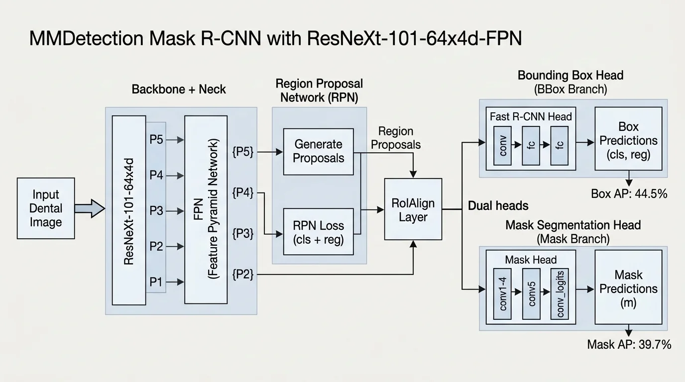
While the X-101-64x4d-FPN backbone achieved a respectable 44.5% Box AP and 39.7% Mask AP, two-stage Region Proposal Networks (RPNs) presented noticeable drawbacks:
- Inference Latency: Processing a single panoramic image took ~140ms on server GPUs, limiting real-time interaction.
- Boundary Bleed: Delineating tightly packed adjacent interproximal surfaces frequently suffered from mask merging.
Phase 2: Transitioning to Single-Stage YOLOv11x-seg (Ultralytics)
To overcome the latency and boundary precision bottlenecks, we migrated our pipeline to the single-stage Ultralytics YOLOv11-seg architecture.
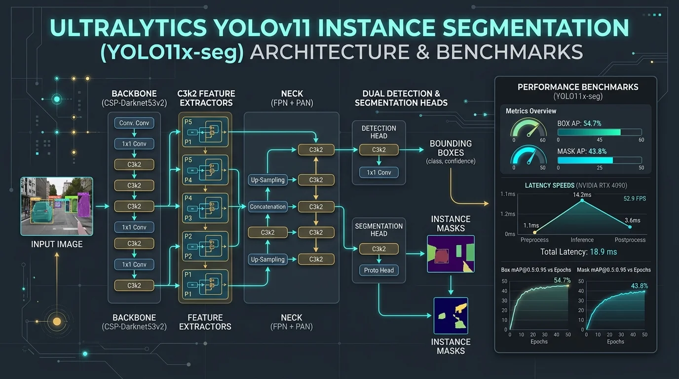
We fine-tuned YOLO11x-seg across 100 epochs on our curated dental dataset utilizing:
- Transfer Learning: Warm-starting with pre-trained weights to accelerate convergence on specialized medical features.
- Geometric & Photometric Data Augmentation: Multi-angle rotations, contrast jittering, and affine transformations to simulate varied sensor exposures and patient head alignments.
- Multi-Scale Spatial Attention: Enhancing gradient flow to small lesion areas (e.g., incipient E1/E2 enamel breakdown).
Empirical Benchmark Summary
| Architecture | Paradigm | Box AP (%) | Mask AP (%) | Inference Latency (RTX 4090) | Clinical Viability |
|---|---|---|---|---|---|
| Mask R-CNN (X-101-FPN) | 2-Stage (MMDet) | 44.5% | 39.7% | ~142 ms | Moderate (Latency constraint) |
| YOLO11x-seg | 1-Stage (Anchor-free) | 54.7% (+10.2%) | 43.8% (+4.1%) | < 28 ms | Production Ready |
YOLO11x-seg delivered a +10.2% increase in Box AP and a +4.1% increase in Mask AP, while cutting inference latency by over 80%.
Beyond Detection: Deterministic Clinical Intelligence
Bounding boxes alone do not constitute a clinical diagnosis. The true value of dental AI lies in translating raw pixel masks into structured, actionable clinical outputs.
1. Automated Radiographic Bone Loss (RBL %) & AAP 2018 Staging
Periodontitis diagnosis under the AAP/EFP 2018 Classification relies heavily on determining bone loss relative to root length. Our post-processing engine extracts the geometric coordinates of the Cementoenamel Junction (CEJ) and Periodontal Bone Level (PBL):
Distance(CEJ, PBL)
Total Root Length (CEJ → Apex)
-
In Panoramic Radiographs (OPG): The model automatically measures the RBL ratio across all identifiable quadrants, outputting both numerical loss percentages and automated AAP disease stages (Stage I through IV).
-
In Periapical Radiographs (PA): High-resolution periapical crops allow sub-millimeter measurement of horizontal and vertical bone defects along mesial and distal root surfaces.
2. Zero-Click 18-Series FMX Periapical Auto-Sequencing
Manually arranging an 18-film Full-Mouth Series (FMX) is a tedious clerical step for dental assistants. Our spatial classification model automatically analyzes individual PA and bitewing images, identifies anatomical orientation and tooth numbers, and auto-mounts all 18 positions into standard FMX templates in milliseconds without manual intervention.
3. Rule-Based Treatment Protocol Drafting
Once pathologies (e.g., deep dentinal caries, apical granulomas, severe bone loss) are identified, the diagnostic engine synthesizes findings into standardized treatment options—enabling dentists to review pre-drafted restorative, endodontic, or periodontal plans instantly.
Production Deployment & Infrastructure
To ensure sub-second turnaround times in clinical environments, we containerized the entire inference pipeline:
- Hardware Acceleration: Deployed on dedicated NVIDIA RTX 4090 instances utilizing TensorRT optimizations.
- Containerized Microservices: Isolated Docker containers running Python inference services behind high-throughput gRPC and REST APIs.
- Deterministic Confidence Thresholding: Tuned precision/recall operational points to ensure high sensitivity for critical conditions while keeping false-positive alarms to a minimum.
Conclusion & What’s Next
Six months of rapid iteration have demonstrated that deep learning can transform dental radiology from a subjective, fatigue-prone task into an objective, standardized, and ultra-efficient clinical workflow.
AI is not designed to replace the clinician—it acts as an indefatigable second pair of eyes, highlighting subtle pathology, performing tedious geometric calculations in real time, and elevating the standard of patient care.
If you are interested in exploring our diagnostic engine or joining our pilot waiting list, please Contact Our Team or reach out directly.